Radiofrequency Ablation
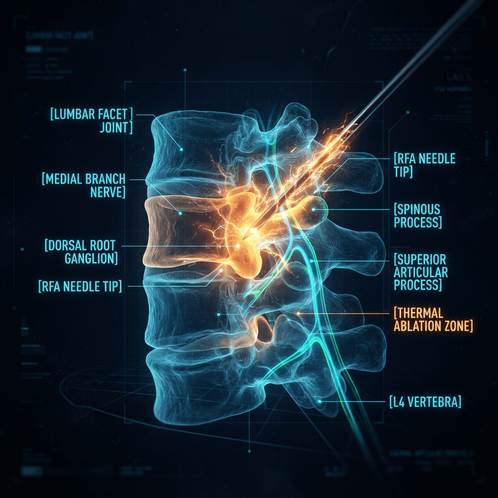
Radiofrequency ablation (RFA) uses heat from radio waves to switch off a specific nerve that carries pain signals. A thin needle is guided to the nerve with live X-ray or ultrasound. It is not surgery: there is no incision, and it is done as an outpatient procedure.
Radiofrequency ablation (RFA) is a minimally invasive procedure that uses heat from radio waves to switch off (disable) a specific nerve that carries pain signals. A physician guides a thin, insulated needle to the target nerve using live X-ray (fluoroscopy) or ultrasound imaging. It is not surgery — it is a needle-based outpatient procedure with no incision.
Key Facts at a Glance
- Most often used for chronic joint-related pain in the back, neck, sacroiliac joint, and knee.
- Relief is long-lasting but not necessarily permanent — the nerve can regenerate over time.
- Also called: radiofrequency neurotomy, rhizotomy, denervation (per ASPN).
- A diagnostic nerve block is usually required before RFA to confirm the nerve is the pain source.
Benefits of Radiofrequency Ablation
RFA targets the nerve carrying the pain signal, often providing longer relief than injections without surgery or a long recovery.
RFA is a proven, minimally invasive option for pain from arthritic joints once a diagnostic block gives temporary relief. Results vary, and relief is not permanent.
- Selected after a positive diagnostic nerve block
- Relief typically lasts months and can be repeated
- Not surgery, and it requires no incision
- Results vary between patients
How Does Radiofrequency Ablation Work?
Pain from an arthritic or irritated joint travels to the brain along tiny sensory nerves. RFA heats a short section of one of these nerves so it can no longer transmit pain signals. Because the targeted nerves carry only sensation from the joint, muscle strength is not meant to change.
The Physics
Radio waves create an electrical field at the needle tip. That field produces heat in the surrounding tissue.
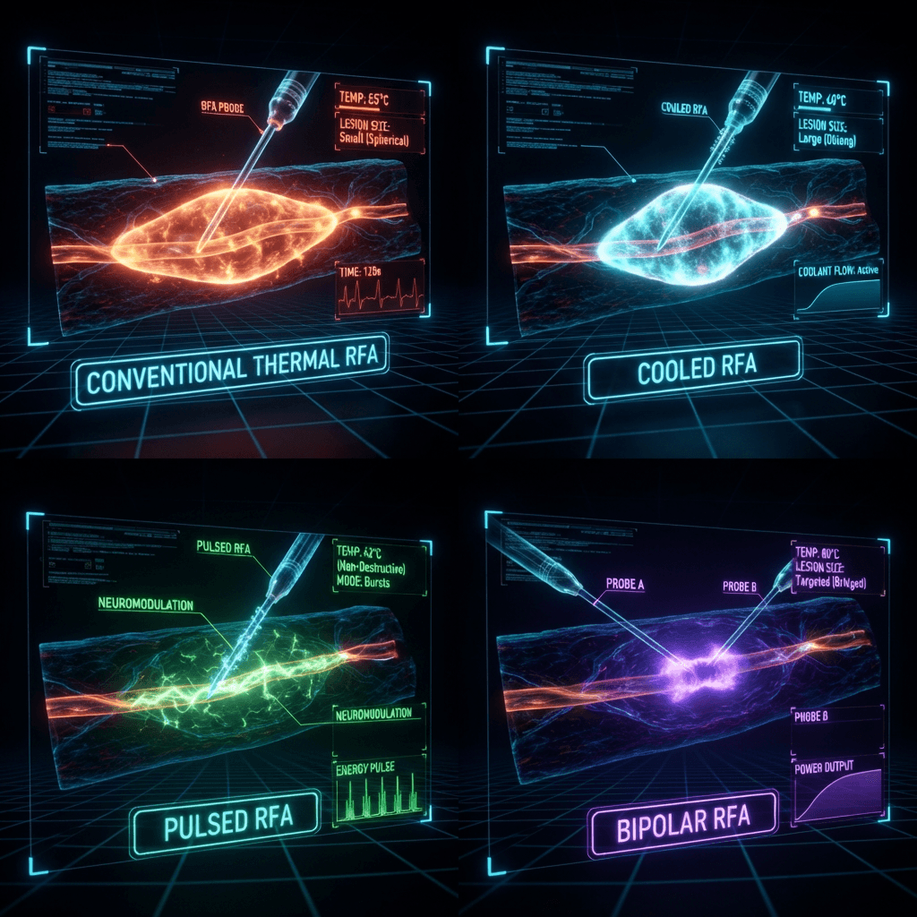
Conventional RFA: The tip reaches a temperature high enough to disrupt (destroy) the nerve.
Pulsed RFA: Energy is delivered in short bursts that keep tissue temperature lower — modulating rather than destroying the nerve.
The goal in every version: Reduce the pain signal reaching the brain.
What Is a Diagnostic Block, and Why Does It Come First?
A diagnostic block is an injection of local anesthetic next to the nerve that RFA would treat. If pain drops meaningfully for the few hours the anesthetic lasts, that nerve is likely carrying the pain. If it does not, RFA is unlikely to help.
Many programs use one or two blocks and set a threshold for relief, such as ≥50% reduction in pain, before proceeding.
Quick Facts
| Question | Short Answer |
|---|---|
| What is it? | Heat energy applied to a pain-carrying (sensory) nerve through a needle |
| What does it treat? | Chronic pain from facet joints, sacroiliac joint, knee, hip, shoulder, and some headaches |
| Is it surgery? | No. It is a needle-based outpatient procedure with no incision |
| Anesthesia | Local anesthetic, with optional light sedation |
| Procedure time | Usually 30–60 minutes per region |
| Recovery | Most people resume light activity within a day or two; soreness at the needle site is common |
| Relief duration | Varies by person and condition; often several months to a year or longer |
| Can it be repeated? | Yes. Nerves can regrow, and repeat procedures are commonly performed |
Types of Radiofrequency Ablation
There are four main types of RFA. The physician chooses based on the target nerve, the anatomy, and the condition being treated. Source: The American Society of Pain and Neuroscience reviews conventional, pulsed and cooled modalities by target area in its LEARN guidelines.
| Type | How It Works | Typical Use | Key Point |
|---|---|---|---|
| Conventional (Thermal) RFA | Continuous energy heats the nerve to create a lesion | Facet joint, sacroiliac joint and many peripheral targets | Most widely used and studied for spinal joint pain |
| Cooled RFA | Water circulates through the needle so a larger lesion can form without overheating the tip | Knee, sacroiliac joint and some spine targets | Larger treatment zone; equipment dependent |
| Pulsed RFA (PRF) | Short bursts keep tissue cooler; modulates rather than destroys the nerve | Neuropathic pain, occipital neuralgia, some shoulder and knee conditions | Non-destructive; evidence is growing (Pain Physician, 2025) |
| Bipolar RFA | Two probes create a lesion between them | Select spine and joint targets | Used in specific anatomy |
Conditions Treated — With Evidence Notes
RFA is most often used for chronic joint-related pain. Each condition has a different evidence base. The best fit and less likely to help notes below are based on the clinical literature cited in this catalogue.

Low Back Pain (Lumbar Facet Joints)
Best Fit
Pain worse with standing, extension, or twisting; felt in the low back and buttocks; without leg weakness.
Less Likely to Help
Pain driven mainly by a pinched nerve (radicular leg pain) or disc problems.
Evidence Note
ASIPP 2026 guideline rates evidence as moderate with a moderate strength of recommendation. The BMJ 2025 guideline recommends against joint RFA in chronic axial spine pain. Both are presented.
Neck Pain (Cervical Facet Joints)
Best Fit
Neck pain with stiffness, worse with turning or looking up, sometimes spreading to the shoulder or base of the skull. Also relevant: whiplash-related facet pain and cervicogenic headache.
Less Likely to Help
Pain driven mainly by a compressed nerve root or disc herniation.
Evidence Note
ASIPP 2026 rates evidence for cervical RFA as moderate with a moderate strength of recommendation.
Sacroiliac (SI) Joint Pain
Best Fit
Pain on one side near the dimples of the low back, worse with standing from sitting, stairs, or rolling in bed.
Less Likely to Help
Pain from disc, hip, or other non-SI sources.
Evidence Note
ASPN's LEARN guidelines address posterior SI joint neurotomy as a distinct target. Cooled and multi-lesion techniques are often used because the nerve supply is variable.
Knee Pain (Genicular Nerve RFA)
Best Fit
Persistent osteoarthritis knee pain, or pain that continues after knee replacement.
Less Likely to Help
Knee pain from causes other than osteoarthritis or post-replacement pain.
Evidence Note
CADTH review noted uncertainty about benefit. Randomized trials of cooled and conventional genicular RFA exist. Many payers still classify genicular RFA as investigational, and coverage varies widely.
Hip Pain
Best Fit
Chronic hip pain, often from arthritis, in people who are poor surgical candidates or who want to delay or avoid hip replacement.
Less Likely to Help
Hip pain from fracture, infection, or instability.
Evidence Note
ASPN's LEARN guidelines cover hip and knee joint neurotomy; evidence is smaller than for spine targets.
Shoulder Pain
Best Fit
Chronic shoulder pain from arthritis, rotator cuff conditions, or after injury when physical therapy and injections have not helped.
Less Likely to Help
Full-thickness rotator cuff tears requiring surgical repair.
Evidence Note
Usually pulsed RFA of the suprascapular nerve. Pulsed RFA has clinical evidence in shoulder pain and knee osteoarthritis (Pain Physician, 2025).
Headache and Occipital Neuralgia
Best Fit
Occipital neuralgia and cervicogenic headache after medication and nerve blocks have given only short-lived relief.
Less Likely to Help
Common migraine or tension headache — RFA is not a first-line treatment for these.
Evidence Note
Clinical evidence supports pulsed RFA in occipital neuralgia, cervicogenic headache and trigeminal neuralgia (Pain Physician, 2025). A neurologic evaluation comes first to rule out other causes.
Mid-Back (Thoracic) Pain
Best Fit
Chronic mid-back pain confirmed by diagnostic blocks after other causes (such as fractures or rib problems) have been excluded.
Less Likely to Help
Mid-back pain from fracture, tumor, or other structural causes.
Evidence Note
ASIPP 2026 reports strong evidence for thoracic RFA, with a weak to moderate strength of recommendation.
Other Uses (Brief Mention)
Pulsed RFA is also reported for: pudendal neuralgia, coccygodynia, meralgia paresthetica, and post-herpetic neuralgia (Pain Physician, 2025).
Who May Be a Good Candidate?
Candidacy Checklist
RFA is considered for adults who have had pain for at least three months, have tried conservative care, and get meaningful temporary relief from a diagnostic nerve block.
- Pain has lasted three months or longer
- Physical therapy, exercise or medication have been tried or are not suitable
- The pain pattern fits a joint or nerve that RFA can treat
- A diagnostic block gave meaningful temporary relief
- No active infection, uncontrolled bleeding risk or other reason to delay
Who May NOT Be a Good Candidate
Active infection
Needle procedures can spread infection
Bleeding disorder or blood thinners that cannot be paused
Higher risk of bleeding
Pain mainly from a pinched nerve root, a fracture, a tumor or an unstable spine
RFA targets joint nerves, not these causes
No relief from a diagnostic block
Suggests the nerve is not the pain source
Pregnancy
Imaging radiation concerns; discuss with physician
Implanted devices (pacemaker, stimulator, intrathecal pump)
Needs coordination; ASIPP's guideline addresses RFA in patients with stimulators and intrathecal systems
Preparation
Medications
Tell your physician about blood thinners, diabetes, infection, pregnancy, and implanted devices such as pacemakers or spinal cord stimulators.
Eating & Drinking
Follow the clinic's instructions on eating, drinking, and medications on the day of the procedure.
Arrange a Ride
Arrange a ride if sedation is planned. Do not drive for the rest of the day if you received sedation.
What to Wear
Wear loose clothing. Clinic-specific details (fasting rules, arrival time, what to bring) should be confirmed by your physician.
What Happens During the Procedure?
Check-in and preparation
Patient changes into a gown; team reviews medications and any blood thinners.
Positioning
Most patients lie face down for spine procedures; position varies by joint.
Skin numbing
Skin is cleaned and numbed with local anesthetic. Light sedation may be offered.
Needle placement
Physician guides the needle to the target nerve using fluoroscopy or ultrasound.
Sensory and motor testing
A small stimulation test confirms position. Patient may feel tingling or muscle twitching in the right location.
Ablation
Energy is delivered for roughly one to three minutes at each site, depending on technique.
Finish
Needle is removed and a small bandage is applied. No stitches are needed.
Monitoring
Patient rests briefly and goes home the same day, usually with a driver if sedation was used.
What It Feels Like
Most people describe pressure and warmth, and some brief achiness. Local anesthetic keeps the heat phase comfortable for most patients.
Recovery Timeline
| When | What to Expect |
|---|---|
| Day of procedure | Rest. Numbness from the anesthetic wears off within hours. Ice packs at the site as directed. |
| Days 1–3 | Soreness, mild bruising or a "sunburn" feeling near the needle sites. Many people return to desk work. |
| Week 1 | Pain may flare temporarily as the nerve settles. Gentle walking is encouraged; follow your activity instructions. |
| Weeks 2–4 | Relief usually becomes noticeable as the nerve effect takes hold. |
| Weeks 4–6 | Follow-up visit to assess response and plan rehabilitation. |
Why a Temporary Flare Is Common
The treated nerve and surrounding tissue are irritated by the procedure. Many patients feel a short-term increase in pain or burning sensation before benefit appears.
How Long Does Relief Last?
Duration varies by condition, technique, and person. Nerves can regrow over months, and pain may return. Often several months to a year or longer. ASIPP 2026 reports similar outcomes with repeat facet interventions.
Important: RFA Does Not Repair the Joint
RFA reduces pain but does not repair the underlying joint. Rehabilitation, strengthening, movement, weight management, and activity pacing remain important.
Opioid Use After RFA
A CADTH review noted evidence that, in chronic axial spine pain, the proportion of patients with opioid prescriptions fell after RFA, while effects for knee, hip and shoulder pain were less clear.
Risks — Common, Uncommon, and Rare
Common and Temporary
- Soreness or bruising at the needle site
- A short-term increase in pain or a burning, sunburn-like feeling
- Numbness or tingling in a small patch of skin
- Temporary fatigue after sedation
Uncommon but Important
- Infection at the needle site
- Bleeding or a hematoma, especially with blood thinners
- Nerve irritation or injury, including temporary weakness
- Skin burn at the treatment site
- Allergic reaction to medication or contrast
- Vascular injury near the knee, because genicular nerves lie close to genicular arteries
A 2025 review describes pulsed RFA as safe and non-ablative with minimal risks (Pain Physician, 2025).
⚠ Warning Signs — Call for Urgent Care If You Have:
Fever, chills, or redness, warmth, swelling or drainage at the needle site
New leg or arm weakness, numbness in the groin or saddle area, or loss of bladder or bowel control
Severe or worsening pain that does not ease with the instructions given
Chest pain, trouble breathing, or signs of an allergic reaction such as hives or swelling of the face
Call emergency services (911 in the US) for emergencies.
Alternatives and How RFA Compares
Nerve ablation does not repair the cause of pain. For many conditions it is used as part of a plan that also includes exercise, weight management and activity pacing.
| Option | What It Does | Typical Role | Trade-offs |
|---|---|---|---|
| Physical therapy and exercise | Builds strength, mobility and function | First-line for most chronic musculoskeletal pain | Needs time and consistency; works well alongside RFA |
| Oral medication (NSAIDs, others) | Reduces inflammation or changes pain processing | Early and ongoing management | Side effects; long-term use needs monitoring |
| Facet or joint injection (steroid and anesthetic) | Anesthetic numbs; steroid may reduce inflammation | Diagnosis and short-term relief | Relief may be brief; the BMJ 2025 guideline recommends against joint injections for chronic axial spine pain |
| Diagnostic medial branch block | Anesthetic on the nerve to test the pain source | Decides whether RFA is likely to help | Relief lasts hours; not itself a long-term treatment |
| Radiofrequency ablation | Disrupts the pain-carrying nerve | After conservative care and a positive block | Relief is not permanent; evidence is debated by guideline panels |
| Pulsed RFA | Modulates nerve activity without destroying it | Neuropathic pain and some peripheral targets | Evidence is growing |
| Epidural steroid injection | Reduces inflammation near a nerve root | Radicular (leg or arm) pain | BMJ 2025 guideline also recommends against epidural injection for chronic radicular pain |
| Spinal cord stimulation | Implanted device sends electrical signals that alter pain perception | Selected refractory pain after other treatments | Surgical implant; needs a trial |
| Surgery (fusion, replacement) | Treats structural problems directly | Instability, severe deformity, end-stage arthritis | Longer recovery; higher procedural risk |
Evidence Summary
The evidence is real but debated. Results are generally better when pain is confirmed to come from the joint by diagnostic blocks before treatment.
Pain Physician 2026 (Manchikanti et al.)
2026 systematic review and meta-analysis of 17 randomized trials rated evidence for RFA in facet joint axial spinal pain as Level II with moderate certainty.
ASIPP June 2026 Guideline
Reports moderate evidence for cervical and lumbar RFA; describes repeat facet interventions as having outcomes similar to the first procedure.
ASPN LEARN Guidelines (2021)
Publishes best-practice guidance by anatomical target (spine to knee and occipital nerves).
BMJ 2025 Guideline (Busse JW et al.)
Made a strong recommendation against joint RFA for non-cancer chronic spine pain, citing very low-certainty evidence. Also recommends against dorsal root ganglion radiofrequency for chronic radicular spine pain and against epidural injection for chronic radicular pain.
Pulsed RFA (Pain Physician, 2025)
A 2025 review describes pulsed RFA as safe, non-ablative, with minimal risks, and with growing evidence (Pain Physician, 2025;28(6):467-481).
CADTH Review
Noted uncertainty about benefit in knee, hip and shoulder joints; noted that the proportion of patients with opioid prescriptions fell after RFA in chronic axial spine pain.
Key Clinical Nuance
Guideline panels differ in how they weigh trial design, patient selection and placebo comparisons. Results are generally better when pain is confirmed to come from the joint by diagnostic blocks before treatment.
Insurance Coverage
| Area | Typical Coverage Picture | Note |
|---|---|---|
| Lumbar, cervical and thoracic facet RFA | Commonly covered with criteria; prior authorization often required | Criteria usually include chronic pain, conservative care, and positive diagnostic blocks |
| Sacroiliac joint RFA | Varies by payer | Verify per plan |
| Genicular (knee) RFA | Often classified as investigational under commercial and Medicare Advantage policies; varies widely | Several payer policies cite a limited or low-quality evidence base; some plans cover it when specific criteria are met |
| Hip and shoulder RFA | Often limited | Verify per plan |
| Medicare | Governed by regional local coverage policies; varies by contractor | Confirm the policy that applies to your region |
Prior Authorization Process
Physician documents pain duration, prior treatments, and examination findings
Diagnostic block results recorded with percentage relief
Clinic submits request to insurer
Approval or denial typically arrives within days to weeks
If denied, you can appeal; the clinic can help with documentation
Frequently Asked Questions
What is radiofrequency ablation for pain?
Radiofrequency ablation (RFA) is a minimally invasive procedure that uses heat from radio waves to disable a pain-carrying nerve. A physician places a thin needle next to the nerve with imaging guidance. It is used for chronic joint and nerve pain, typically after other treatments have not given enough relief.
Is radiofrequency ablation the same as rhizotomy or neurotomy?
Yes, in everyday use. Radiofrequency neurotomy, rhizotomy, denervation and RFA all describe using radiofrequency energy to treat a nerve. ASPN notes that radiofrequency neurotomy is also called radiofrequency ablation.
Is radiofrequency ablation surgery?
No. It is a needle-based outpatient procedure with no incision, usually done with local anesthetic and sometimes light sedation.
Does radiofrequency ablation burn the nerve?
It heats a small section of the nerve to disrupt pain signaling. In pulsed RFA, short energy bursts keep temperatures lower and change nerve activity without destroying tissue. Either way, the goal is pain reduction, not damage to surrounding structures.
What conditions does radiofrequency ablation treat?
RFA is most often used for chronic pain from the facet joints of the neck, mid-back and low back, the sacroiliac joint, and the knee. It is also used for some hip, shoulder and occipital nerve problems.
Who is a good candidate for radiofrequency ablation?
Adults with chronic pain of at least three months who have tried conservative care and who get meaningful temporary relief from a diagnostic nerve block are generally good candidates. The physician makes the final decision after an examination and review of imaging.
Can RFA help with sciatica or a pinched nerve?
RFA generally treats pain from joints, not from a compressed nerve root. Radicular leg pain usually needs a different approach. A 2025 BMJ guideline recommends against dorsal root ganglion radiofrequency for chronic radicular spine pain.
Does radiofrequency ablation work for knee pain?
Some patients with knee osteoarthritis report relief from genicular nerve RFA, but the evidence is less established than for spine targets, and insurance coverage is inconsistent. A diagnostic genicular nerve block usually comes first.
Can RFA treat headaches?
Pulsed radiofrequency has clinical evidence in occipital neuralgia, cervicogenic headache and trigeminal neuralgia (Pain Physician). It is not a first-line treatment for common migraine or tension headache.
How long does a radiofrequency ablation take?
Most procedures take about 30 to 60 minutes, depending on how many nerves are treated. Add time for check-in and recovery.
Does radiofrequency ablation hurt?
Most patients feel pressure, warmth or brief achiness. Local anesthetic numbs the skin and deeper tissues, and sedation may be available. Soreness afterward is common and usually temporary.
Will I be awake during the procedure?
Usually yes, so you can tell the physician what you feel during testing. Light sedation can be offered to keep you relaxed.
Do I need someone to drive me home?
Yes, if you receive sedation. Without sedation, ask your clinic for driving instructions.
How long is recovery after radiofrequency ablation?
Most people return to light activity in one to two days. Soreness at the needle site can last several days, and a temporary pain flare is common before relief builds over two to four weeks.
How long does pain relief last after RFA?
Often several months to a year or longer, but it varies. Nerves can regrow, so pain may return. The procedure can be repeated, and the ASIPP 2026 guideline reports similar outcomes with repeat facet interventions.
How many times can you have RFA?
There is no fixed lifetime limit. Insurers often set frequency limits such as a minimum interval between treatments.
What if radiofrequency ablation doesn't work?
The physician will review whether the right nerve was targeted and whether another source is driving the pain. Other options include different injections, medication changes, physical therapy, or referral for other procedures.
Is radiofrequency ablation safe?
RFA is generally low-risk. Common effects are temporary soreness and bruising. Serious complications such as infection, bleeding or nerve injury are uncommon.
Is radiofrequency ablation proven to work?
Evidence varies by condition and guideline. A 2026 meta-analysis of 17 randomized trials found moderate-certainty evidence for facet joint RFA (Pain Physician), while a 2025 BMJ guideline recommended against joint RFA for chronic spine pain based on very low-certainty evidence.
Does insurance cover radiofrequency ablation?
Often yes for facet joint RFA when criteria are met, with prior authorization. Coverage for knee, hip and shoulder RFA varies widely, and many plans treat genicular RFA as investigational.
How much does radiofrequency ablation cost?
Cost depends on the area treated, the facility and your insurance. Factors include: body area and number of levels/nerves treated; type of RFA (conventional, cooled or pulsed); facility (office-based, ambulatory surgery center, or hospital outpatient); sedation and imaging used; number of diagnostic blocks before treatment; the plan's deductible, copay and coinsurance.
Glossary of RFA and Pain Terms
| Term | Plain-Language Definition |
|---|---|
| Radiofrequency ablation (RFA) | A procedure that uses radio-wave heat to disrupt a nerve that carries pain signals |
| Radiofrequency neurotomy / rhizotomy | Other names for RFA applied to a nerve |
| Facet joints | Small paired joints along the back of the spine that guide movement and can become arthritic and painful |
| Medial branch nerves | Tiny nerves that carry pain signals from the facet joints |
| Medial branch block | A diagnostic injection of local anesthetic that tests whether a medial branch nerve is the pain source |
| Sacroiliac (SI) joint | The joint where the spine meets the pelvis |
| Genicular nerves | Sensory nerves around the knee that carry pain from the joint |
| Occipital nerves | Nerves at the back of the head that can cause occipital neuralgia |
| Conventional (thermal) RFA | Continuous radiofrequency energy that heats and disrupts the nerve |
| Cooled RFA | RFA with water cooling through the needle to create a larger lesion |
| Pulsed RFA (PRF) | Short bursts of energy that modulate a nerve without heat destruction |
| Fluoroscopy | Live X-ray imaging used to guide the needle |
| Lesion | The small, controlled area of nerve tissue affected by the treatment |
| Radicular pain | Pain caused by an irritated or compressed nerve root, often radiating into an arm or leg |
| Axial pain | Pain felt in the spine itself (neck, mid-back or low back) |
| Neuropathic pain | Pain caused by damage or dysfunction in the nervous system |
| Prior authorization | Insurer approval required before a procedure |
| Local coverage determination (LCD) | A regional Medicare policy on what is covered |
| Cervicogenic headache | Headache that starts in the neck structures |
| Occipital neuralgia | Sharp, shooting pain in the back of the head from irritation of the occipital nerves |
Questions to Ask at Your Consultation
Which structure do you think is causing my pain, and how will we confirm it?
What relief should I expect and for how long?
What are the risks for my situation?
What if it does not help?
How many procedures do you expect I will need over time?
Sources
Manchikanti L, et al. Effectiveness of Radiofrequency Ablation in Managing Chronic Axial Spinal Pain of Facet Joint Origin: A Systematic Review and Meta-Analysis of Randomized Controlled Trials. Pain Physician, 2026;29(3):205–240.
ASIPP. Facet Joint Interventions in the Management of Chronic Spinal Pain (guideline, June 2026).
Busse JW, et al. Commonly used interventional procedures for non-cancer chronic spine pain: a clinical practice guideline. BMJ, 2025;388:e079970.
Lee DW, et al. Latest Evidence-Based Application for Radiofrequency Neurotomy (LEARN): Best Practice Guidelines from ASPN. J Pain Res, 2021;14:2807–2831.
Pain Physician. Pulsed Radiofrequency Treatment: Evidence for and Applications in Chronic Pain. 2025;28(6):467–481.
Idahor CO, Okoye J, et al. Radiofrequency Ablation for Chronic Pain: Mechanistic Insights and Emerging Innovations. Cureus, 2025;17(12):e99056.
CADTH. Radiofrequency Ablation for Chronic Knee, Hip, and Shoulder Pain.
This page is for educational purposes only and does not constitute individualized medical advice. Results vary. All evidence, credentials, prices, contact details and claims must be clinically verified before publishing.
Ready to Discuss Radiofrequency Ablation?
A diagnostic evaluation is the first step. Our team will review your history, imaging, and prior treatments to determine whether RFA may be appropriate for your situation.